| 書目名稱 | Magnetic Resonance Imaging of Central Nervous System Diseases |
| 副標題 | Functional Anatomy — |
| 編輯 | W. J. Huk,G. Gademann,G. Friedmann |
| 視頻video | http://file.papertrans.cn/622/621328/621328.mp4 |
| 圖書封面 |  |
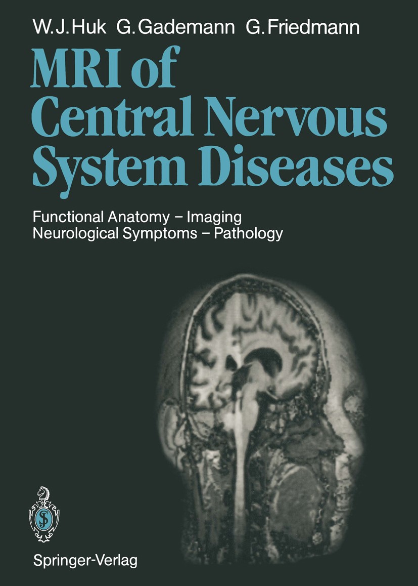
| 描述 | Magnetic resonance imaging (MRI) is a new and still rapidly developing imaging technique which requires a new approach to image interpreta- tion. Radiologists are compelled to translate their experience accumulat- ed from X-ray techniques into the language of MRI, and likewise stu- dents of radiology and interested clinicians need special training in both languages. Out of this necessity emerged the concept of this book as a manual on the application and evaluation of proton MRI for the radiolo- gist and as a guide for the referring physician who wants to learn about the diagnostic value of MRI in specific conditions. After a short section on the basic principles of MRI, the contrast mechanisms of present-day imaging techniques, knowledge of which is essential for the analysis of relaxation times, are described in greater de- tail. This is followed by a demonstration of functional neuroanatomy us- ing three-dimensional view of MR images and a synopsis of frequent neurological symptoms and their topographic correlations, which will fa- cilitate examination strategy with respect to both accurate diagnosis and economy. |
| 出版日期 | Book 1990 |
| 關鍵詞 | Nervous System; Tumor; brainstem; classification; magnetic resonance imaging (MRI); radiotherapy |
| 版次 | 1 |
| doi | https://doi.org/10.1007/978-3-642-72568-5 |
| isbn_softcover | 978-3-642-72570-8 |
| isbn_ebook | 978-3-642-72568-5 |
| copyright | Springer-Verlag Berlin Heidelberg 1990 |
 |Archiver|手機版|小黑屋|
派博傳思國際
( 京公網(wǎng)安備110108008328)
GMT+8, 2025-10-6 03:53


