找回密碼
 To register

QQ登錄

只需一步,快速開始

掃一掃,訪問微社區(qū)

打印 上一主題 下一主題

Titlebook: Atlas of Pathologic Myopia; Kyoko Ohno-Matsui Book 2020 Springer Nature Singapore Pte Ltd. 2020 Pathologic Myopia.Optical coherence tomogr

[復(fù)制鏈接]
查看: 45998|回復(fù): 53
樓主
發(fā)表于 2025-3-21 16:46:53 | 只看該作者 |倒序?yàn)g覽 |閱讀模式
期刊全稱Atlas of Pathologic Myopia
影響因子2023Kyoko Ohno-Matsui
視頻videohttp://file.papertrans.cn/165/164408/164408.mp4
發(fā)行地址Features a wealth of images from the world’s largest high myopia clinic.Images were obtained with state-of-the-art technologies.Includes treatment indications and outcomes with numerous case series.Vi
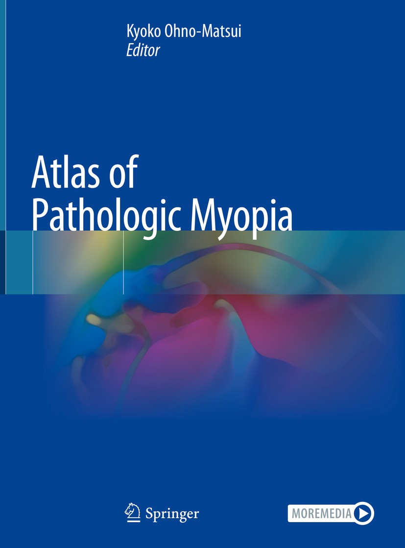
圖書封面Titlebook: Atlas of Pathologic Myopia;  Kyoko Ohno-Matsui Book 2020 Springer Nature Singapore Pte Ltd. 2020 Pathologic Myopia.Optical coherence tomogr
影響因子This Atlas provides many beautiful images obtained with state-of-the-art technologies, including optical coherence tomography (OCT), OCT angiography, fundus autofluorescence, and wide-field fundus imaging, as well as traditional images and fluorescein/ICG angiograms. Gathered at the world’s largest High Myopia Clinic, the images are based on the long-term follow-up data of more than 6,000 patients from Japan and abroad.?Recent advances in imaging technologies have yielded many new observations and allowed us to detect new lesions, e.g. myopic traction maculopathy (or macular retinoschisis) and dome-shaped macula. An especially interesting aspect: the images obtained by ‘3D MRI of the eye’ and ‘ultra wide-field OCT’ to visualize staphylomas. These techniques were established by the editor’s group and make it possible to record the entire shapes of the eye, offering a scan width of up to 23 mm and scan depth of 5 mm. They have since been used to visualize posterior staphyloma,which was previously impossible to view because it spanned such a wide range of the eye. In addition, readers will learn what types of eye deformity occur in pathologic myopia and how they damage the macula/opti
Pindex Book 2020
The information of publication is updating

書目名稱Atlas of Pathologic Myopia影響因子(影響力)


書目名稱Atlas of Pathologic Myopia影響因子(影響力)學(xué)科排名


書目名稱Atlas of Pathologic Myopia網(wǎng)絡(luò)公開度


書目名稱Atlas of Pathologic Myopia網(wǎng)絡(luò)公開度學(xué)科排名


書目名稱Atlas of Pathologic Myopia被引頻次


書目名稱Atlas of Pathologic Myopia被引頻次學(xué)科排名


書目名稱Atlas of Pathologic Myopia年度引用


書目名稱Atlas of Pathologic Myopia年度引用學(xué)科排名


書目名稱Atlas of Pathologic Myopia讀者反饋


書目名稱Atlas of Pathologic Myopia讀者反饋學(xué)科排名


單選投票, 共有 1 人參與投票
 

0票 0.00%

Perfect with Aesthetics


0票 0.00%

Better Implies Difficulty


1票 100.00%

Good and Satisfactory


0票 0.00%

Adverse Performance


0票 0.00%

Disdainful Garbage

您所在的用戶組沒有投票權(quán)限
沙發(fā)
發(fā)表于 2025-3-21 23:22:13 | 只看該作者
板凳
發(fā)表于 2025-3-22 04:03:44 | 只看該作者
TMDU Classification and Curtin’s ClassificationIt is highly characteristic and specific to pathologic myopia since it rarely occurs except for pathologic myopia. The most commonly used classification is the Curtin’s classification, which was based on his observations of the fundi by an ophthalmoscope. Using Optos and 3D MRI, Ohno-Matsui proposed
地板
發(fā)表于 2025-3-22 05:18:48 | 只看該作者
Ultra-Wide Field OCT of Posterior Staphylomaor staphylomas shown by UWF-OCT are gradual thinning of the choroid from the periphery toward the edge of the staphyloma and a gradual re-thickening of the choroid in the direction toward the posterior pole along with a posterior displacement of the sclera in the staphylomatous area. Also, there is
5#
發(fā)表于 2025-3-22 08:56:28 | 只看該作者
6#
發(fā)表于 2025-3-22 14:09:41 | 只看該作者
Macular Diffuse Choroidal Atrophy and eventually extends to the macula. OCT shows a marked thinning of the choroid in the area of diffuse atrophy. In some cases, almost the entire choroid is lost leaving sporadically remaining large choroidal vessels.
7#
發(fā)表于 2025-3-22 17:49:56 | 只看該作者
Patchy Choroidal Atrophynally oval, or round, or irregular when multiple lesions merge. Patchy atrophy can derive from lacquer cracks or diffuse atrophy. It rarely affects the central fovea. Fundus autofluorescence shows uniform hypo-autofluorescence in the area of patchy atrophy. OCT images show a loss of retinal pigment
8#
發(fā)表于 2025-3-23 00:43:28 | 只看該作者
Myopic Macular Neovascularization (Diagnosis)d into three phases: active, scar, and atrophic phase. MNV tends to be smaller and less active than choroidal neovascularization due to age-related macular degeneration. In addition, the presence of background choroidal atrophy makes the detection of MNV difficult in some cases. Thus, multimodal ima
9#
發(fā)表于 2025-3-23 01:22:06 | 只看該作者
10#
發(fā)表于 2025-3-23 08:40:22 | 只看該作者
Kyoko Ohno-MatsuiFeatures a wealth of images from the world’s largest high myopia clinic.Images were obtained with state-of-the-art technologies.Includes treatment indications and outcomes with numerous case series.Vi
 關(guān)于派博傳思  派博傳思旗下網(wǎng)站  友情鏈接
派博傳思介紹 公司地理位置 論文服務(wù)流程 影響因子官網(wǎng) 吾愛論文網(wǎng) 大講堂 北京大學(xué) Oxford Uni. Harvard Uni.
發(fā)展歷史沿革 期刊點(diǎn)評(píng) 投稿經(jīng)驗(yàn)總結(jié) SCIENCEGARD IMPACTFACTOR 派博系數(shù) 清華大學(xué) Yale Uni. Stanford Uni.
QQ|Archiver|手機(jī)版|小黑屋| 派博傳思國(guó)際 ( 京公網(wǎng)安備110108008328) GMT+8, 2025-10-19 07:51
Copyright © 2001-2015 派博傳思   京公網(wǎng)安備110108008328 版權(quán)所有 All rights reserved
快速回復(fù) 返回頂部 返回列表
遵义县| 镇原县| 吉隆县| 泰宁县| 新乐市| 宿迁市| 合川市| 肥乡县| 衡东县| 壶关县| 阿鲁科尔沁旗| 沙洋县| 大新县| 翁源县| 石门县| 盐边县| 沙洋县| 伊吾县| 蒙阴县| 博野县| 保山市| 临澧县| 怀来县| 通化县| 昭苏县| 宁化县| 甘谷县| 肇源县| 大渡口区| 林口县| 六安市| 乌兰浩特市| 广宁县| 昌宁县| 夏河县| 邢台市| 山丹县| 仁布县| 鹿邑县| 汨罗市| 新沂市|