標題: Titlebook: Cutaneous Atlas of Ex Vivo Confocal Microscopy; Manu Jain,Anthony Rossi,Mercedes Sendín-Martín Book 2022 The Editor(s) (if applicable) and [打印本頁] 作者: Ingrown-Toenail 時間: 2025-3-21 18:50
書目名稱Cutaneous Atlas of Ex Vivo Confocal Microscopy影響因子(影響力)
書目名稱Cutaneous Atlas of Ex Vivo Confocal Microscopy影響因子(影響力)學(xué)科排名
書目名稱Cutaneous Atlas of Ex Vivo Confocal Microscopy網(wǎng)絡(luò)公開度
書目名稱Cutaneous Atlas of Ex Vivo Confocal Microscopy網(wǎng)絡(luò)公開度學(xué)科排名
書目名稱Cutaneous Atlas of Ex Vivo Confocal Microscopy被引頻次
書目名稱Cutaneous Atlas of Ex Vivo Confocal Microscopy被引頻次學(xué)科排名
書目名稱Cutaneous Atlas of Ex Vivo Confocal Microscopy年度引用
書目名稱Cutaneous Atlas of Ex Vivo Confocal Microscopy年度引用學(xué)科排名
書目名稱Cutaneous Atlas of Ex Vivo Confocal Microscopy讀者反饋
書目名稱Cutaneous Atlas of Ex Vivo Confocal Microscopy讀者反饋學(xué)科排名
作者: Carminative 時間: 2025-3-21 20:48
http://image.papertrans.cn/d/image/241655.jpg作者: 山崩 時間: 2025-3-22 04:01
https://doi.org/10.1007/978-3-030-75035-0on-neoplastic lesions, enabling bedside pathology. Ex vivoconfocal microscopy (EVCM) acquires images in two modes: reflectance confocal microscopy (RCM) and fluorescence confocal microscopy (FCM). RCM (grayscale) and FCM (grayscale or green scale) images can be visualized separately or in a combined作者: Presbyopia 時間: 2025-3-22 05:37
https://doi.org/10.1007/978-3-030-75035-0w generations of EVCM microscopes combine two different simultaneous diode lasers (reflectance and fluorescence), to create a fusion pseudo-colored image of the scanned tissue called Fusion confocal microscopy (fuCM) with digital H&E (DHE), as it mimics conventionally stained hematoxylin and eosin (作者: Customary 時間: 2025-3-22 10:43
Sequences and Series of Functions, is now being integrated during Mohs surgery for margin assessment of the residual skin cancers, such as basal cell carcinoma (BCC) and squamous cell carcinoma (SCC). However, to identify skin cancers it is quintessential to first learn the features of normal skin on EVCME. Using this device, fresh 作者: CONE 時間: 2025-3-22 14:32
The Structure Of Systems Analysiss (SK) are benign nonmelanocytic epidermal lesions that are commonly seen on sun-damaged skin of patients undergoing Mohs surgery; thus, it is crucial to identify them on frozen sections or freshly excised tissues and differentiate them from skin cancers such as basal cell carcinoma (BCCC), squamous作者: CONE 時間: 2025-3-22 19:14
The Structure Of Systems Analysis, schwannoma), fibrous (dermatofibroma, acrochordon), adnexal (sebaceous hyperplasia), or mesenchymal (lipoma, angiolipoma) origin. Although benign in nature, these lesions often clinically mimic malignant lesions such as BCC or other adnexal tumors, requiring biopsy for histopathology confirmation.作者: 神經(jīng) 時間: 2025-3-22 23:05 作者: 火海 時間: 2025-3-23 01:39
Views on Art, Tragedy, and Fiction,-rapid diagnostic tool may be extended beyond non-melanoma skin cancer, especially basal cell carcinoma, to other indications including melanocytic lesions. Current knowledge and experience on the use of EVCM in melanocytic lesions are limited but shows promising horizons. Examples of junctional, co作者: Recessive 時間: 2025-3-23 09:29 作者: instate 時間: 2025-3-23 12:20
https://doi.org/10.1057/978-1-137-58897-5 to safely distinguish normal or dysplastic nevi from melanomaintraoperatively and immediately decide further therapeutic steps would potentially decrease the number of surgicalprocedures, as well as associated risk of complications. Possible use of ex vivo confocal laser scanning microscopy (ex viv作者: Affectation 時間: 2025-3-23 15:15 作者: 興奮過度 時間: 2025-3-23 19:44 作者: VAN 時間: 2025-3-24 00:58
Dysplastic Neviaser scanning microscopy (ex vivo CLSM). The innovative bedside examination of freshly excised tissue has previously been applied also on melanocytic lesions. We present current knowledge and images of dysplastic melanocytic nevi studied with ex vivo CLSM.作者: 媒介 時間: 2025-3-24 04:27 作者: enlist 時間: 2025-3-24 08:18
A Handbook of Tropical PaediatricsThe availability of an instantaneous histological diagnosis through the use of ex vivo confocal microscopy (evCM) has completely changed pathology analysis not only of the skin but also in general pathology.作者: antidepressant 時間: 2025-3-24 12:19 作者: TRAWL 時間: 2025-3-24 17:09
Squamous Cell Carcinoma Features on Ex Vivo Confocal Imaging and Histopathologic CorrelationThe availability of an instantaneous histological diagnosis through the use of ex vivo confocal microscopy (evCM) has completely changed pathology analysis not only of the skin but also in general pathology.作者: 小故事 時間: 2025-3-24 21:56 作者: Ringworm 時間: 2025-3-25 02:45
978-3-030-89318-7The Editor(s) (if applicable) and The Author(s), under exclusive license to Springer Nature Switzerl作者: Inflated 時間: 2025-3-25 04:13
https://doi.org/10.1057/978-1-137-58897-5aser scanning microscopy (ex vivo CLSM). The innovative bedside examination of freshly excised tissue has previously been applied also on melanocytic lesions. We present current knowledge and images of dysplastic melanocytic nevi studied with ex vivo CLSM.作者: Heart-Rate 時間: 2025-3-25 08:02 作者: 混雜人 時間: 2025-3-25 14:11 作者: Duodenitis 時間: 2025-3-25 19:18 作者: AMBI 時間: 2025-3-25 21:48 作者: 黃油沒有 時間: 2025-3-26 01:26
Features of Benign Nonmelanocytic Dermal and Subcutaneous Lesions on Ex Vivo Confocal Microscopy and, schwannoma), fibrous (dermatofibroma, acrochordon), adnexal (sebaceous hyperplasia), or mesenchymal (lipoma, angiolipoma) origin. Although benign in nature, these lesions often clinically mimic malignant lesions such as BCC or other adnexal tumors, requiring biopsy for histopathology confirmation.作者: 先兆 時間: 2025-3-26 08:18 作者: Judicious 時間: 2025-3-26 10:19 作者: antedate 時間: 2025-3-26 15:52 作者: Gleason-score 時間: 2025-3-26 18:27 作者: 美色花錢 時間: 2025-3-26 23:29
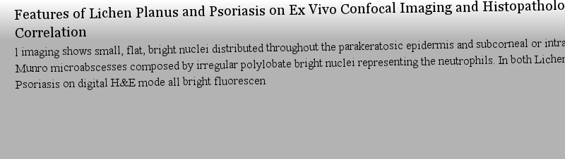
Features of Lichen Planus and Psoriasis on Ex Vivo Confocal Imaging and Histopathologic Correlationes and histologically featured by a dense, band-like lymphocytic infiltrate and keratinocyte apoptosis with destruction of the epidermal basal cell layer. Ex vivo confocal imaging shows dense band of bright small and round nuclei distributed in the dermis immediately below the epidermis. Psoriasis i作者: sterilization 時間: 2025-3-27 02:29
Eczema: Features on FCM, Digital H&E, and Corresponding Conventional H&Ethe dynamism of the inflammatory sequencies occurring in the epidermis and dermal portion and include epidermal hyperplasia, ortho-parakeratosis, spongiosis, and inflammatory cells accumulation in the dermis. Ex vivo fluorescent confocal images show crowded bright nuclei of hyperplastic epidermis an作者: Commemorate 時間: 2025-3-27 08:03
ice, especially in Mohs surgery for the evaluation of keratinocytic neoplasm and in dermatopathology for rapid evaluation of?varied skin lesions. It is therefore a valuable resource for trainee, residents, prac978-3-030-89318-7978-3-030-89316-3作者: ADORE 時間: 2025-3-27 12:03
https://doi.org/10.1007/978-3-030-75035-0eneration EVCM, Vivascope 2500 (Caliber ID, Rochester, NY, USA) and the most commonly used fluorescent dye, acridine orange. The technique detailed here is based on the methods described by the experts in the literature and on our personal experience of imaging and handling tissues in dermatology re作者: 脫水 時間: 2025-3-27 17:34 作者: Harbor 時間: 2025-3-27 21:06 作者: CLAN 時間: 2025-3-27 22:12 作者: Confound 時間: 2025-3-28 03:21
https://doi.org/10.1057/9780230236998l imaging shows small, flat, bright nuclei distributed throughout the parakeratosic epidermis and subcorneal or intracorneal Munro microabscesses composed by irregular polylobate bright nuclei representing the neutrophils. In both Lichen Planus and Psoriasis on digital H&E mode all bright fluorescen作者: 懸崖 時間: 2025-3-28 09:35
Book 2022x Vivo Confocal Microscopy.?covers how to apply these techniques into dermatological practice, especially in Mohs surgery for the evaluation of keratinocytic neoplasm and in dermatopathology for rapid evaluation of?varied skin lesions. It is therefore a valuable resource for trainee, residents, prac作者: Phenothiazines 時間: 2025-3-28 13:27 作者: 小臼 時間: 2025-3-28 16:27 作者: adumbrate 時間: 2025-3-28 22:30
Features of Benign Epidermal Nonmelanocytic Lesions on Ex Vivo Confocal Microscopy and Histopathologd SK have never been described in the literature. In this chapter, we will first provide a brief introduction to clinical and pathologic features of SL, SK, VV, and MC and then define morphological features of these lesions on EVCM (fluorescence and digital H&E modes) and compare them with their his作者: impaction 時間: 2025-3-29 01:44 作者: 誘拐 時間: 2025-3-29 04:48 作者: Microaneurysm 時間: 2025-3-29 09:43
Morphological Features of Basal Cell Carcinoma on Ex Vivo Confocal Imaging and Histopathologic Correng tumor margin assessment in real-time. EVCM has shown high sensitivity and specificity for detecting BCCs. In this chapter, we have described the morphological features of common subtypes of BCC as visualized in various imaging modes on the EVCM (fluorescence confocal microscopy and digital H&E mode) and compared them with their corresponding.作者: cardiopulmonary 時間: 2025-3-29 14:37
MelanomaoCLSM) in melanoma diagnostics, including fast immunofluorescence and intraoperative tumor thickness measurement, aswell as up-to-date experience together with examples of melanoma images are presented and discussed.作者: 的事物 時間: 2025-3-29 16:30
Eczema: Features on FCM, Digital H&E, and Corresponding Conventional H&Ed bright nuclei of lymphocytes in the epidermal-dermal junction. Dark spaces in the basal layer of the epidermis are consistent with spongiosis. On digital H&E mode the bright white structures on FCM appear dark purple-blue, corresponding to hyperchromatic hematoxylin stained nuclei on histopathology.作者: Enthralling 時間: 2025-3-29 21:40
https://doi.org/10.1007/978-3-030-75035-0hematoxylin stain) and signal from RCM (cytoplasmic and collagen signal) which is digitally converted to pink color (similar to eosin stain). For information about the principles of ECM device, please refer to Chap. ..作者: 尾隨 時間: 2025-3-30 03:18
Roles and Applications of Ex Vivo Confocal Microscopyhematoxylin stain) and signal from RCM (cytoplasmic and collagen signal) which is digitally converted to pink color (similar to eosin stain). For information about the principles of ECM device, please refer to Chap. ..作者: 小溪 時間: 2025-3-30 05:13 作者: 錯誤 時間: 2025-3-30 11:48
Acquired Nevi: Junctional, Compound, and Dermalsions. Current knowledge and experience on the use of EVCM in melanocytic lesions are limited but shows promising horizons. Examples of junctional, compound, and dermal nevi with classic EVCMEx vivo confocal microscopy features are presented in this chapter.作者: harangue 時間: 2025-3-30 15:23
https://doi.org/10.1007/978-94-011-5907-4ng tumor margin assessment in real-time. EVCM has shown high sensitivity and specificity for detecting BCCs. In this chapter, we have described the morphological features of common subtypes of BCC as visualized in various imaging modes on the EVCM (fluorescence confocal microscopy and digital H&E mode) and compared them with their corresponding.作者: VEST 時間: 2025-3-30 17:57
https://doi.org/10.1057/978-1-137-58897-5oCLSM) in melanoma diagnostics, including fast immunofluorescence and intraoperative tumor thickness measurement, aswell as up-to-date experience together with examples of melanoma images are presented and discussed.作者: overwrought 時間: 2025-3-30 23:53
https://doi.org/10.1057/9780230236998d bright nuclei of lymphocytes in the epidermal-dermal junction. Dark spaces in the basal layer of the epidermis are consistent with spongiosis. On digital H&E mode the bright white structures on FCM appear dark purple-blue, corresponding to hyperchromatic hematoxylin stained nuclei on histopathology.作者: Mumble 時間: 2025-3-31 04:51 作者: PRO 時間: 2025-3-31 08:18
Views on Art, Tragedy, and Fiction,sions. Current knowledge and experience on the use of EVCM in melanocytic lesions are limited but shows promising horizons. Examples of junctional, compound, and dermal nevi with classic EVCMEx vivo confocal microscopy features are presented in this chapter.