標題: Titlebook: Clinical Anatomy of the Head; Neurocranium · Orbit Johannes Lang Book 1983 Springer-Verlag Berlin Heidelberg 1983 Hirnsch?del.Kraniozervika [打印本頁] 作者: Awkward 時間: 2025-3-21 16:49
書目名稱Clinical Anatomy of the Head影響因子(影響力)
書目名稱Clinical Anatomy of the Head影響因子(影響力)學(xué)科排名
書目名稱Clinical Anatomy of the Head網(wǎng)絡(luò)公開度
書目名稱Clinical Anatomy of the Head網(wǎng)絡(luò)公開度學(xué)科排名
書目名稱Clinical Anatomy of the Head被引頻次
書目名稱Clinical Anatomy of the Head被引頻次學(xué)科排名
書目名稱Clinical Anatomy of the Head年度引用
書目名稱Clinical Anatomy of the Head年度引用學(xué)科排名
書目名稱Clinical Anatomy of the Head讀者反饋
書目名稱Clinical Anatomy of the Head讀者反饋學(xué)科排名
作者: 腫塊 時間: 2025-3-21 22:06 作者: 高興去去 時間: 2025-3-22 00:51 作者: 傻 時間: 2025-3-22 06:53
Synopsis of the Skull,Various points and lines have been established by anatomists, anthropologists and clinicians as aids to studying the growth and dimensions of the skull and its normal and abnormal shapes.作者: abject 時間: 2025-3-22 11:54 作者: Cleave 時間: 2025-3-22 15:15
Midbrain and Adjacent Structures,The mesencephalon connects those parts of the brain in the hypophyseal region (diencephalon) with those in the posterior cranial fossa (rhombencephalon).作者: Cleave 時間: 2025-3-22 20:44
http://image.papertrans.cn/c/image/227726.jpg作者: nonchalance 時間: 2025-3-22 21:14 作者: bioavailability 時間: 2025-3-23 02:12 作者: BACLE 時間: 2025-3-23 06:57 作者: conservative 時間: 2025-3-23 09:48 作者: calumniate 時間: 2025-3-23 16:48 作者: legislate 時間: 2025-3-23 21:37 作者: 磨碎 時間: 2025-3-24 00:50 作者: 根除 時間: 2025-3-24 06:01
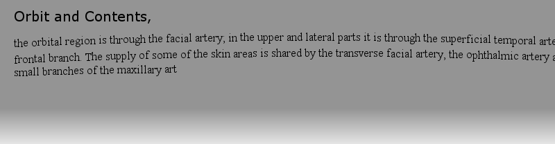
https://doi.org/10.1007/978-3-319-31361-0the orbital region is through the facial artery; in the upper and lateral parts it is through the superficial temporal artery and its frontal branch. The supply of some of the skin areas is shared by the transverse facial artery, the ophthalmic artery and numerous small branches of the maxillary art作者: forager 時間: 2025-3-24 09:27
https://doi.org/10.1007/978-3-319-44947-0to the posterolateral part (L. and B. 1981). 21. The thinnest zones of the floor of the anterior cranial fossa average 0.2 mm in the newborn and increase in thickness with age, though very inconsistently, reaching 0.66–1.13 mm at 17 years of age.作者: Madrigal 時間: 2025-3-24 11:11 作者: 碎石頭 時間: 2025-3-24 17:56 作者: 提名的名單 時間: 2025-3-24 19:36 作者: canvass 時間: 2025-3-25 00:31
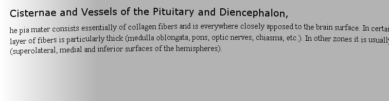
Loan Guarantees Schemes in Risk Management,he pia mater consists essentially of collagen fibers and is everywhere closely apposed to the brain surface. In certain regions this layer of fibers is particularly thick (medulla oblongata, pons, optic nerves, chiasma, etc.). In other zones it is usually thinner (superolateral, medial and inferior 作者: Flavouring 時間: 2025-3-25 06:56 作者: tenuous 時間: 2025-3-25 08:25 作者: 徹底檢查 時間: 2025-3-25 15:20
Risk Classification in Life Insuranceher to the transverse sinus or the sigmoid sinus. Less commonly they run medially to the superior petrosal sinus or the straight sinus. At their origins the sinuses are usually 3–4 mm in diameter, broadening near their terminations to 8–10 mm. In length they vary between 3 and 73 mm. The termination作者: LITHE 時間: 2025-3-25 16:55 作者: 哭得清醒了 時間: 2025-3-25 21:06 作者: 娘娘腔 時間: 2025-3-26 01:41
https://doi.org/10.1007/978-3-319-44947-0to the posterolateral part (L. and B. 1981). 21. The thinnest zones of the floor of the anterior cranial fossa average 0.2 mm in the newborn and increase in thickness with age, though very inconsistently, reaching 0.66–1.13 mm at 17 years of age.作者: 熱情的我 時間: 2025-3-26 05:12
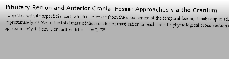
Chitra Rajagopal,Indra Deo Kumar. Together with its superficial part, which also arises from the deep lamina of the temporal fascia, it makes up in adults approximately 37.5% of the total mass of the muscles of mastication on each side. Its physiological cross-section amounts to approximately 4.1 cm.. For further details see L./W., Praktische Anatomie, Vol. I, 1C.作者: 輕率的你 時間: 2025-3-26 08:56 作者: 后來 時間: 2025-3-26 14:41 作者: commune 時間: 2025-3-26 19:21 作者: fatuity 時間: 2025-3-26 22:50 作者: 遺留之物 時間: 2025-3-27 03:32 作者: 寡頭政治 時間: 2025-3-27 05:26 作者: 迷住 時間: 2025-3-27 09:35
Posterior Cranial Fossa and Contents, of those which run into the transverse sinus is situated an average of 50 (10–80) mm from the confluence of the sinuses (Fig. 266). In very rare cases we found a longitudinal tentorial sinus (see Fig. 265).作者: 乳汁 時間: 2025-3-27 15:35
https://doi.org/10.1007/978-3-319-44947-0or is bounded by the inferior orbital fissure while anterolaterally it curves upwards into the lateral wall of the orbit. The medial region of the floor is somewhat elevated and merges smoothly into the medial wall of the orbit.作者: 不能平靜 時間: 2025-3-27 19:25
Chitra Rajagopal,Indra Deo Kumarary lobe, where it frequently divides into two branches which embrace the posterior lobe. A ramus superior runs on the upper posterior part of the posterior lobe, and a ramus inferior on the lower posterior part (Fig. 131).作者: mighty 時間: 2025-3-28 01:29 作者: Maximizer 時間: 2025-3-28 03:04 作者: Misgiving 時間: 2025-3-28 10:15
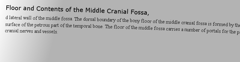
Results of the empirical studies,d lateral wall of the middle fossa. The dorsal boundary of the bony floor of the middle cranial fossa is formed by the anterior surface of the petrous part of the temporal bone. The floor of the middle fossa carries a number of portals for the passage of cranial nerves and vessels.作者: left-ventricle 時間: 2025-3-28 14:12 作者: countenance 時間: 2025-3-28 16:51
Risk Classification in Life Insurance of those which run into the transverse sinus is situated an average of 50 (10–80) mm from the confluence of the sinuses (Fig. 266). In very rare cases we found a longitudinal tentorial sinus (see Fig. 265).作者: 植物學(xué) 時間: 2025-3-28 19:59
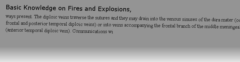
Diploic Veins, Meninges and Scalp,ways present. The diploic veins traverse the sutures and they may drain into the venous sinuses of the dura mater (occipital, frontal and posterior temporal diploic veins) or into veins accompanying the frontal branch of the middle meningeal artery (anterior temporal diploic vein). Communications wi作者: GRACE 時間: 2025-3-29 00:02 作者: scrape 時間: 2025-3-29 05:57
Anterior Cranial Fossa, the Approach to the Orbit and the Ethmoid Bone,to the posterolateral part (L. and B. 1981). 21. The thinnest zones of the floor of the anterior cranial fossa average 0.2 mm in the newborn and increase in thickness with age, though very inconsistently, reaching 0.66–1.13 mm at 17 years of age.作者: lattice 時間: 2025-3-29 10:04
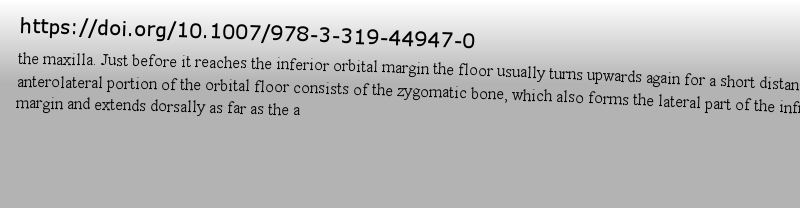
The Floor of the Orbit,the maxilla. Just before it reaches the inferior orbital margin the floor usually turns upwards again for a short distance. The anterolateral portion of the orbital floor consists of the zygomatic bone, which also forms the lateral part of the infraorbital margin and extends dorsally as far as the a作者: Mettle 時間: 2025-3-29 14:42 作者: 有效 時間: 2025-3-29 16:29 作者: BILE 時間: 2025-3-29 21:49 作者: Condyle 時間: 2025-3-30 00:52 作者: construct 時間: 2025-3-30 04:12 作者: Feigned 時間: 2025-3-30 10:39 作者: 不能仁慈 時間: 2025-3-30 15:14
Craniocervical Junction,ally, two ex-occipital or lateral occipital centers follow on each side immediately adjacent to the hypoglossal canals, around which bone will eventually grow downwards. The condylar parts of the occipital bone ossify from the basi-occipital and lateral occipital centers. A pair of supra-occipital o作者: Dysplasia 時間: 2025-3-30 19:23 作者: commensurate 時間: 2025-3-30 22:20
Book 1983e skull base upwards, but any such plan would have required a photoatlas in several volumes. For this reason I have confined myself to medical problems of current importance. In this volume I have included numerous variations which I have myself encountered, so as to underline the diversity of human作者: Prologue 時間: 2025-3-31 04:14
ad from the skull base upwards, but any such plan would have required a photoatlas in several volumes. For this reason I have confined myself to medical problems of current importance. In this volume I have included numerous variations which I have myself encountered, so as to underline the diversity of human978-3-642-68242-1作者: ineptitude 時間: 2025-3-31 08:29 作者: 異端邪說2 時間: 2025-3-31 12:44 作者: Reclaim 時間: 2025-3-31 13:54
Craniocervical Junction,onnected by wide bridges of cartilage, the anterior intraoccipital synchondroses. The squamous part of the occipital bone is connected to the lateral parts by the posterior intraoccipital synchondroses.作者: scotoma 時間: 2025-3-31 17:48