標題: Titlebook: Atlas of Advanced Endoscopy; Yasushi Sano,Philip Chiu,Chikatoshi Katada Book 2024 The Editor(s) (if applicable) and The Author(s), under e [打印本頁] 作者: 租期 時間: 2025-3-21 16:43
書目名稱Atlas of Advanced Endoscopy影響因子(影響力)
書目名稱Atlas of Advanced Endoscopy影響因子(影響力)學(xué)科排名
書目名稱Atlas of Advanced Endoscopy網(wǎng)絡(luò)公開度
書目名稱Atlas of Advanced Endoscopy網(wǎng)絡(luò)公開度學(xué)科排名
書目名稱Atlas of Advanced Endoscopy被引頻次
書目名稱Atlas of Advanced Endoscopy被引頻次學(xué)科排名
書目名稱Atlas of Advanced Endoscopy年度引用
書目名稱Atlas of Advanced Endoscopy年度引用學(xué)科排名
書目名稱Atlas of Advanced Endoscopy讀者反饋
書目名稱Atlas of Advanced Endoscopy讀者反饋學(xué)科排名
作者: maverick 時間: 2025-3-21 21:34
Classification of Endoscopic Imaginghereafter various fiberscopes were also applied clinically. In 1984, electronic videoendoscopes were also developed. Unlike fiberscopes, which directly detect light signals, electronic videoendoscopes convert electronic signals into images via semiconductor elements and allow various forms of electronic image processing and analysis.作者: frozen-shoulder 時間: 2025-3-22 01:43
How to Take High-Quality Images (H&N)ts in magnified endoscopy, numerous cases of squamous cell carcinoma are now being detected through gastrointestinal endoscopy. In the detection and characterization of squamous cell carcinoma, NBI magnification (M-NBI) is significantly superior to white light endoscopy (WLE), making M-NBI an essential tool in daily clinical practice [1, 2].作者: Ringworm 時間: 2025-3-22 05:03 作者: 結(jié)果 時間: 2025-3-22 09:09
Pharyngeal PapillomaSquamous papilloma, pharynx.作者: 使聲音降低 時間: 2025-3-22 13:24
https://doi.org/10.1007/978-981-97-2732-2Endoscopy; Classification; IEE; Image-enhanced endoscopy; Gastrointestinal tract; Cancer作者: Modicum 時間: 2025-3-22 18:06
978-981-97-2734-6The Editor(s) (if applicable) and The Author(s), under exclusive license to Springer Nature Singapor作者: 閑蕩 時間: 2025-3-22 21:30
Theo Drane,M. V. Achutha Kiran Kumar an image enhancement technique that forms contrast of deep tissue and blood vessels and is expected to be used more widely in the future. In this chapter, we will describe the principle of RDI and its clinical applications.作者: indices 時間: 2025-3-23 02:07
The Enhanced Entity-Relationship Modelhereafter various fiberscopes were also applied clinically. In 1984, electronic videoendoscopes were also developed. Unlike fiberscopes, which directly detect light signals, electronic videoendoscopes convert electronic signals into images via semiconductor elements and allow various forms of electronic image processing and analysis.作者: Confirm 時間: 2025-3-23 07:34
Handbook of Condition Monitoringts in magnified endoscopy, numerous cases of squamous cell carcinoma are now being detected through gastrointestinal endoscopy. In the detection and characterization of squamous cell carcinoma, NBI magnification (M-NBI) is significantly superior to white light endoscopy (WLE), making M-NBI an essential tool in daily clinical practice [1, 2].作者: Pelvic-Floor 時間: 2025-3-23 12:36 作者: 大酒杯 時間: 2025-3-23 15:06
http://image.papertrans.cn/b/image/167640.jpg作者: OVER 時間: 2025-3-23 20:59 作者: Nostalgia 時間: 2025-3-24 00:16
Springer Professional Computing visibility of lesions by improving three vital image factors inherent to white light imaging (WLI): (1) brightness in dark areas; (2) texture, such as subtle surface changes; and (3) color, including slight color variations in the image. To achieve this, TXI applies image processing technology base作者: bleach 時間: 2025-3-24 03:37
Theo Drane,M. V. Achutha Kiran Kumar an image enhancement technique that forms contrast of deep tissue and blood vessels and is expected to be used more widely in the future. In this chapter, we will describe the principle of RDI and its clinical applications.作者: 香料 時間: 2025-3-24 08:59
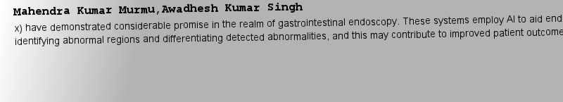
Mahendra Kumar Murmu,Awadhesh Kumar Singhnt focus has been the integration of AI in medical imaging. In particular, systems referred to as computer-aided detection and diagnosis (CADe and CADx) have demonstrated considerable promise in the realm of gastrointestinal endoscopy. These systems employ AI to aid endoscopists in identifying abnor作者: 小卷發(fā) 時間: 2025-3-24 11:31 作者: Orchiectomy 時間: 2025-3-24 16:06 作者: Budget 時間: 2025-3-24 20:18
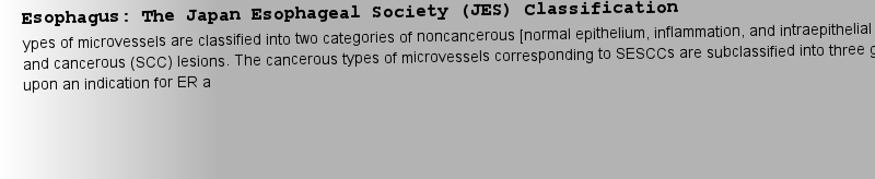
Meike Klettke,Bernhard Thalheim(ER) because the rate of lymph node metastasis increases in proportion to the invasion depth of the carcinoma. Previous studies have shown a close relationship between microvascular patterns observed by magnifying endoscopy and invasion depth of the superficial carcinoma. Although there were two maj作者: 極端的正確性 時間: 2025-3-25 01:05
Handbook of Conceptual Modeling(EAC), of which the 5-year overall survival rate is a dismal 17% [1, 2]. International guidelines have recommended endoscopic surveillance for patients with BE to detect early dysplasia [3, 4]. Endoscopic surveillance is typically performed with high-definition white light imaging (WLI) with quadran作者: linguistics 時間: 2025-3-25 06:06
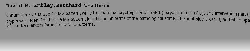
David W. Embley,Bernhard Thalheim(M-NBI) of the stomach [2]. The VS classification system employs anatomical terms as endoscopic markers for analysis. Anatomical components visualized using M-NBI were divided into microvascular (MV) and microsurface (MS) patterns. As shown in ? Fig. 1a–c, the subepithelial capillary and collecting 作者: 運動的我 時間: 2025-3-25 08:48
Meike Klettke,Bernhard Thalheimcation [1]. NBI observation became widely recognized as an essential method for colorectal tumor diagnosis. In 2009, an international collaborative research group called the Colon Tumor NBI Interest Group (CTNIG), led by experts from Japan (Yasushi Sano, Shinji Tanaka), France (Ponchon T), the Unite作者: Volatile-Oils 時間: 2025-3-25 12:30 作者: MEN 時間: 2025-3-25 18:22 作者: 財主 時間: 2025-3-25 20:08 作者: 雜色 時間: 2025-3-26 02:46
EVIS X1 Endoscopy Systemcal signals and displaying them on a monitor was developed in Japan in 1986 [1]. Subsequently, models such as CV-240 (1997), CV-260, Lucera Spectrum (2006), and CV-290, Lucera Elite (2010) have evolved remarkably in terms of image quality and functionality. Notably, the recent introduction of the CV作者: 伙伴 時間: 2025-3-26 07:06 作者: Defense 時間: 2025-3-26 10:54 作者: 壓迫 時間: 2025-3-26 13:10
Artificial Intelligence (AI) in Colonoscopynt focus has been the integration of AI in medical imaging. In particular, systems referred to as computer-aided detection and diagnosis (CADe and CADx) have demonstrated considerable promise in the realm of gastrointestinal endoscopy. These systems employ AI to aid endoscopists in identifying abnor作者: jealousy 時間: 2025-3-26 19:03 作者: cajole 時間: 2025-3-26 22:56
Paris Classificationor “superficial” neoplastic lesions in the esophagus, stomach, and colon [1]. A neoplastic lesion is considered “superficial” when its endoscopic appearance suggests that it has not penetrated the digestive wall beyond the submucosa. In Japan, neoplastic stomach lesions that appear “superficial” dur作者: hardheaded 時間: 2025-3-27 04:16 作者: MOT 時間: 2025-3-27 06:18
Esophagus: The BING Classification(EAC), of which the 5-year overall survival rate is a dismal 17% [1, 2]. International guidelines have recommended endoscopic surveillance for patients with BE to detect early dysplasia [3, 4]. Endoscopic surveillance is typically performed with high-definition white light imaging (WLI) with quadran作者: 態(tài)度暖昧 時間: 2025-3-27 13:29
Stomach: VS Classification/MESDA-G(M-NBI) of the stomach [2]. The VS classification system employs anatomical terms as endoscopic markers for analysis. Anatomical components visualized using M-NBI were divided into microvascular (MV) and microsurface (MS) patterns. As shown in ? Fig. 1a–c, the subepithelial capillary and collecting 作者: liaison 時間: 2025-3-27 16:14
Colon: The Japan NBI Expert Team (JNET) Classificationcation [1]. NBI observation became widely recognized as an essential method for colorectal tumor diagnosis. In 2009, an international collaborative research group called the Colon Tumor NBI Interest Group (CTNIG), led by experts from Japan (Yasushi Sano, Shinji Tanaka), France (Ponchon T), the Unite作者: Albumin 時間: 2025-3-27 21:27
How to Take High-Quality Images (H&N)ts in magnified endoscopy, numerous cases of squamous cell carcinoma are now being detected through gastrointestinal endoscopy. In the detection and characterization of squamous cell carcinoma, NBI magnification (M-NBI) is significantly superior to white light endoscopy (WLE), making M-NBI an essent作者: Homocystinuria 時間: 2025-3-27 22:15 作者: Perennial長期的 時間: 2025-3-28 03:38
How to Take High-Quality Images (LGI)ehensive, and clear to reflect endoscopic findings and interventions faithfully [1]. Moreover, it is the best method to verify the completeness of an endoscopic procedure. Therefore, photo documentation is an essential aspect of endoscopic quality control. In addition, taking high-quality endoscopic作者: 憲法沒有 時間: 2025-3-28 09:52
Book 2024fication, the VS Classification/MESDA-G, and the JNET Classification. Each case section provides expert analysis and notable endoscopic images, accompanied by histopathologic images depicting various benign lesions and early cancers...The Atlas of Advanced Endoscopy. serves as an invaluable resource作者: Legend 時間: 2025-3-28 13:22 作者: Evocative 時間: 2025-3-28 16:29 作者: 抱狗不敢前 時間: 2025-3-28 21:52 作者: HACK 時間: 2025-3-28 23:55
Meike Klettke,Bernhard Thalheimed to provide more accurate differential diagnoses, linking appropriate treatment strategies. Therefore, the Japan NBI Expert Team (JNET) conducted a web-based prospective study to validate the magnifying NBI findings (principal investigator: Yasushi Sano) and finally proposed the JNET classification in 2014 (? Fig. 1) [2, 3].作者: 有偏見 時間: 2025-3-29 04:44
Common vibration monitoring techniquesfying colonoscopy observation are also skilled in techniques such as EMR/ESD. In the future, AI diagnosis may become widespread, but endoscopists need to capture appropriate and high-quality images to use AI assistance effectively. For the novice endoscopist, image documentation is the first step toward becoming an expert endoscopist.作者: Cubicle 時間: 2025-3-29 09:38
Colon: The Japan NBI Expert Team (JNET) Classificationed to provide more accurate differential diagnoses, linking appropriate treatment strategies. Therefore, the Japan NBI Expert Team (JNET) conducted a web-based prospective study to validate the magnifying NBI findings (principal investigator: Yasushi Sano) and finally proposed the JNET classification in 2014 (? Fig. 1) [2, 3].作者: Parallel 時間: 2025-3-29 13:47
How to Take High-Quality Images (LGI)fying colonoscopy observation are also skilled in techniques such as EMR/ESD. In the future, AI diagnosis may become widespread, but endoscopists need to capture appropriate and high-quality images to use AI assistance effectively. For the novice endoscopist, image documentation is the first step toward becoming an expert endoscopist.作者: 桶去微染 時間: 2025-3-29 18:28 作者: 梯田 時間: 2025-3-29 21:01
Anargyros Sarafopoulos,Bernard F. Buxton-1500, EVIS X1 (2020), marks a significant milestone after a decade and heralds further advancements (? Fig. 1). ? Table 1 provides a summary of the distinctions between the previous and the latest models.作者: CLAY 時間: 2025-3-30 03:17
Springer Professional Computingd on the retinex theory [1]. This theory involves decomposing images into two layers based on human visual system characteristics [2]. The first layer represents the scene’s illumination light, while the second layer corresponds to the local contrast in brightness and color.作者: engender 時間: 2025-3-30 07:21 作者: ABIDE 時間: 2025-3-30 08:34
Commercial applications of visual monitoring analyze the information for research, and (3) with readers worldwide if published in a medical journal. High-quality endoscopic images are better beautiful, but they must be informative and narrative. In this sense, endoscopic photodocumentation is similar to photojournalism.作者: remission 時間: 2025-3-30 15:11 作者: 剝削 時間: 2025-3-30 20:03 作者: Blazon 時間: 2025-3-30 23:07
Handbook of Conceptual Modelings with BE to detect early dysplasia [3, 4]. Endoscopic surveillance is typically performed with high-definition white light imaging (WLI) with quadrantic biopsies taken as per the Seattle protocol [5]. However, early Barrett’s dysplasia are easily missed due to subtle mucosal change [6, 7].作者: 殺菌劑 時間: 2025-3-31 02:18
Artificial Intelligence (AI) in Colonoscopyx) have demonstrated considerable promise in the realm of gastrointestinal endoscopy. These systems employ AI to aid endoscopists in identifying abnormal regions and differentiating detected abnormalities, and this may contribute to improved patient outcomes.作者: Anthrp 時間: 2025-3-31 05:38
Esophagus: The BING Classifications with BE to detect early dysplasia [3, 4]. Endoscopic surveillance is typically performed with high-definition white light imaging (WLI) with quadrantic biopsies taken as per the Seattle protocol [5]. However, early Barrett’s dysplasia are easily missed due to subtle mucosal change [6, 7].