標(biāo)題: Titlebook: Atlas of Parathyroid Imaging and Pathology; Alexander L. Shifrin,L. Daniel Neistadt,Pritinder Book 2020 The Editor(s) (if applicable) and [打印本頁(yè)] 作者: Amalgam 時(shí)間: 2025-3-21 17:00
書目名稱Atlas of Parathyroid Imaging and Pathology影響因子(影響力)
書目名稱Atlas of Parathyroid Imaging and Pathology影響因子(影響力)學(xué)科排名
書目名稱Atlas of Parathyroid Imaging and Pathology網(wǎng)絡(luò)公開(kāi)度
書目名稱Atlas of Parathyroid Imaging and Pathology網(wǎng)絡(luò)公開(kāi)度學(xué)科排名
書目名稱Atlas of Parathyroid Imaging and Pathology被引頻次
書目名稱Atlas of Parathyroid Imaging and Pathology被引頻次學(xué)科排名
書目名稱Atlas of Parathyroid Imaging and Pathology年度引用
書目名稱Atlas of Parathyroid Imaging and Pathology年度引用學(xué)科排名
書目名稱Atlas of Parathyroid Imaging and Pathology讀者反饋
書目名稱Atlas of Parathyroid Imaging and Pathology讀者反饋學(xué)科排名
作者: 兩棲動(dòng)物 時(shí)間: 2025-3-21 20:19
Functions of Completely Regular Growth different protocols. Some protocols use both 60 and 90 second runs (3 post contrast phases) which may be helpful when rate of opacification is not optimal. The parathyroid gland or adenoma washes out in the venous phase, distinguishing it from enhancing nodes, which usually have no washout or incre作者: animated 時(shí)間: 2025-3-22 02:13
Pathology of the Parathyroid Glandsrlying hyperparathyroidism, including parathyroid adenoma, atypical parathyroid adenoma, parathyroid hyperplasia, and parathyroid carcinoma. Syndromes of familial hyperparathyroidism are cataloged in the context of their molecular pathogenesis. Less common lesions covered include parathyroid cyst, p作者: Promotion 時(shí)間: 2025-3-22 05:07
Individual CT Phases different protocols. Some protocols use both 60 and 90 second runs (3 post contrast phases) which may be helpful when rate of opacification is not optimal. The parathyroid gland or adenoma washes out in the venous phase, distinguishing it from enhancing nodes, which usually have no washout or incre作者: 擁護(hù) 時(shí)間: 2025-3-22 11:32
parathyroid pathology is also included to help the reader understand challenges in pathological interpretation...Atlas of Parathyroid Imaging and Pathology. serves as a valuable reference for radiologists, end978-3-030-40961-6978-3-030-40959-3作者: indoctrinate 時(shí)間: 2025-3-22 15:34 作者: compose 時(shí)間: 2025-3-22 17:13
A toolkit for several dimensionss that are 1–2 mm thick. The fourth dimension is enhancement change over time. Axial, sagittal, and coronal sections 1 mm thick are generated from all the runs. Parathyroid tissue has prominent arterial phase enhancement and good venous washout.作者: 單片眼鏡 時(shí)間: 2025-3-22 21:42 作者: tympanometry 時(shí)間: 2025-3-23 04:13 作者: 使激動(dòng) 時(shí)間: 2025-3-23 09:21
Fonctions d‘une variable réellezation of a parathyroid adenoma helps the surgeon to perform a minimally invasive parathyroidectomy with a targeted approach to a single-gland parathyroid adenoma, rather than performing neck exploration of all four parathyroid glands. Surgeon-performed ultrasound detects parathyroid adenoma more of作者: foreign 時(shí)間: 2025-3-23 13:39
Fonctions d‘une variable réelleonomously functioning adenoma, hyperplasia, and rarely, carcinoma. Parathyroidectomy is highly effective in achieving durable biochemical cure. Distinguishing single-gland adenoma and multigland hyperplasia is aided by preoperative imaging localization studies. Intraoperative assessment includes fro作者: affluent 時(shí)間: 2025-3-23 14:56
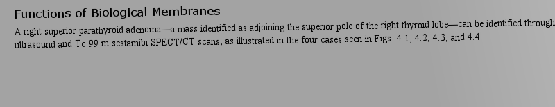
https://doi.org/10.1007/978-1-4899-3252-5ocated posteriorly and inferiorly to the inferior pole of the right thyroid lobe. When Tc99m sestamibi SPECT/CT scans are used, the scintigraphic findings are compatible with a parathyroid adenoma at the level of the lower pole of the right lobe of the thyroid gland. On immediate images a focus of i作者: Androgen 時(shí)間: 2025-3-23 18:48
Experimental approaches to mechanism,riorly to the superior pole of the left thyroid lobe. When sestamibi scans are used, in some cases persistent tracer activity within each lobe of the thyroid gland severely limits evaluation for a parathyroid adenoma, even on 3-hour delayed images. When viewed in light of the ultrasound findings, ho作者: GRE 時(shí)間: 2025-3-24 00:27 作者: 攤位 時(shí)間: 2025-3-24 05:22 作者: Annotate 時(shí)間: 2025-3-24 09:09
Integrability of the Fourier transforms on arteriography and prominent enhancement on CTscan. Multiple studies have shown excellant contrast CT scan detection of enlarged parathyroid glands and adenomas as initial screening study and in cases of failed sestamibi studies. The hypervascularity is also identifiable on color Doppler ultrasou作者: 五行打油詩(shī) 時(shí)間: 2025-3-24 12:09
A toolkit for several dimensionss that are 1–2 mm thick. The fourth dimension is enhancement change over time. Axial, sagittal, and coronal sections 1 mm thick are generated from all the runs. Parathyroid tissue has prominent arterial phase enhancement and good venous washout.作者: HEPA-filter 時(shí)間: 2025-3-24 18:40 作者: endocardium 時(shí)間: 2025-3-24 22:04 作者: eczema 時(shí)間: 2025-3-25 01:41
https://doi.org/10.1007/978-94-011-2418-8oid glands and parathyroid adenomas are usually predominantly echo-poor relative to the thyroid, and vascularity is usually demonstrated on color Doppler. Though the parathyroid adenoma is typically diffusely echo-poor (at least 50% of the time), echogenic components may be seen within the enlarged 作者: 協(xié)迫 時(shí)間: 2025-3-25 04:16
The CT Techniques that are 1–2 mm thick. The fourth dimension is enhancement change over time. Axial, sagittal, and coronal sections 1 mm thick are generated from all the runs. Parathyroid tissue has prominent arterial phase enhancement and good venous washout.作者: rectum 時(shí)間: 2025-3-25 07:54 作者: 克制 時(shí)間: 2025-3-25 12:04
http://image.papertrans.cn/b/image/164406.jpg作者: 首創(chuàng)精神 時(shí)間: 2025-3-25 18:26
https://doi.org/10.1007/978-3-540-34038-6Parathyroid scintigraphy has been used for the detection and preoperative planning of primary hyperthyroidism. A variety of scintigraphic imaging techniques can be used for preoperative localization.作者: seroma 時(shí)間: 2025-3-25 22:38 作者: 類似思想 時(shí)間: 2025-3-26 02:24
https://doi.org/10.1007/978-1-4899-3252-5A left inferior parathyroid adenoma—a mass identified as adjoining the inferior pole of the left thyroid lobe—can be identified through imaging with ultrasound and Tc 99 m sestamibi SPECT/CT scans, as illustrated in the four cases seen in Figs. 7.1, 7.2, 7.3, and 7.4.作者: 閑逛 時(shí)間: 2025-3-26 04:52
Properties of the membrane phase,This chapter presents eight cases of intrathyroidal parathyroid adenoma (Figs. 8.1, 8.2, 8.3, 8.4, 8.5, and 8.6) or cystic parathyroid adenoma (Figs. 8.7 and 8.8), which are all identified using ultrasound and SPECT/CT imaging.作者: 黃油沒(méi)有 時(shí)間: 2025-3-26 10:53
Scintigraphic Parathyroid ImagingParathyroid scintigraphy has been used for the detection and preoperative planning of primary hyperthyroidism. A variety of scintigraphic imaging techniques can be used for preoperative localization.作者: 女上癮 時(shí)間: 2025-3-26 14:15 作者: LOPE 時(shí)間: 2025-3-26 18:21 作者: GIDDY 時(shí)間: 2025-3-26 21:42
Ultrasonography, Sestamibi Scan, and SPECT/CT Sestamibi Scan of Intrathyroidal Parathyroid Adenoma aThis chapter presents eight cases of intrathyroidal parathyroid adenoma (Figs. 8.1, 8.2, 8.3, 8.4, 8.5, and 8.6) or cystic parathyroid adenoma (Figs. 8.7 and 8.8), which are all identified using ultrasound and SPECT/CT imaging.作者: inflate 時(shí)間: 2025-3-27 04:31 作者: 棲息地 時(shí)間: 2025-3-27 06:44 作者: Spongy-Bone 時(shí)間: 2025-3-27 09:38
Right Inferior Parathyroid Adenomaocated posteriorly and inferiorly to the inferior pole of the right thyroid lobe. When Tc99m sestamibi SPECT/CT scans are used, the scintigraphic findings are compatible with a parathyroid adenoma at the level of the lower pole of the right lobe of the thyroid gland. On immediate images a focus of i作者: 同步信息 時(shí)間: 2025-3-27 15:58
Left Superior Parathyroid Adenomariorly to the superior pole of the left thyroid lobe. When sestamibi scans are used, in some cases persistent tracer activity within each lobe of the thyroid gland severely limits evaluation for a parathyroid adenoma, even on 3-hour delayed images. When viewed in light of the ultrasound findings, ho作者: debris 時(shí)間: 2025-3-27 19:33
Imaging of the Parathyroid Carcinomaoidism. Because of the rarity of this cancer, the diagnosis is difficult to establish. The diagnosis could be suspected from findings of a very high serum calcium level usually higher than 14–16?mg/dL with corresponding elevation of serum parathyroid hormone level 10–15-fold higher than the normal r作者: TOM 時(shí)間: 2025-3-27 23:48 作者: prosthesis 時(shí)間: 2025-3-28 03:18 作者: 敲竹杠 時(shí)間: 2025-3-28 07:44 作者: instructive 時(shí)間: 2025-3-28 12:08
Individual CT Phaseslarly important for distinguishing subcapsular parathyroid glands, but it is also critical in identifying ectopic rests of thyroid tissue. Arterial phase is performed during contrast infusion that starts 20–45 seconds from the start of a rapid 3 or 4 cc/second infusion of contrast followed by a sali作者: 小爭(zhēng)吵 時(shí)間: 2025-3-28 17:33 作者: 拍下盜公款 時(shí)間: 2025-3-28 20:57
Correlative Ultrasoundoid glands and parathyroid adenomas are usually predominantly echo-poor relative to the thyroid, and vascularity is usually demonstrated on color Doppler. Though the parathyroid adenoma is typically diffusely echo-poor (at least 50% of the time), echogenic components may be seen within the enlarged 作者: figment 時(shí)間: 2025-3-29 00:26 作者: Regurgitation 時(shí)間: 2025-3-29 04:29 作者: cathartic 時(shí)間: 2025-3-29 08:53
Experimental approaches to mechanism,wever, a very mild persistent focus of increased tracer activity may be identified. More accurate testing to detect a parathyroid adenoma would consist of a SPECT/CT sestamibi scan (rather than a regular sestamibi scan) in combination with a dedicated parathyroid ultrasound study or 4D CT scan.作者: 首創(chuàng)精神 時(shí)間: 2025-3-29 12:24 作者: Ganglion 時(shí)間: 2025-3-29 19:10 作者: 工作 時(shí)間: 2025-3-29 19:50 作者: 揭穿真相 時(shí)間: 2025-3-30 03:37 作者: 支柱 時(shí)間: 2025-3-30 07:59
Imaging of the Parathyroid Carcinoma that can clearly establish the diagnosis of parathyroid carcinoma, but some imaging modalities such as ultrasound and CT scan can be helpful in the initial evaluation of patients with a suspected diagnosis of parathyroid carcinoma. Sestamibi scans can also help in localization.作者: 鑲嵌細(xì)工 時(shí)間: 2025-3-30 11:35 作者: 顛簸地移動(dòng) 時(shí)間: 2025-3-30 15:18 作者: 輕率的你 時(shí)間: 2025-3-30 16:34
Motivation for Imaging Studiestinal surgery is performed before a neck exploration. Ultrasound, sestamibi scan, contrast CT scan, and MRI are the noninvasive modalities available to find the starting point in evaluating patients with clinically diagnosed primary hyperparathyroidism.作者: CRATE 時(shí)間: 2025-3-30 21:53 作者: Indecisive 時(shí)間: 2025-3-31 03:51 作者: CARE 時(shí)間: 2025-3-31 08:13