標(biāo)題: Titlebook: Atlas of Echocardiography in Pediatrics and Congenital Heart Diseases; Maryam Moradian,Azin Alizadehasl Book 2021 Springer-Verlag GmbH Ger [打印本頁(yè)] 作者: 解毒藥 時(shí)間: 2025-3-21 17:35
書(shū)目名稱(chēng)Atlas of Echocardiography in Pediatrics and Congenital Heart Diseases影響因子(影響力)
書(shū)目名稱(chēng)Atlas of Echocardiography in Pediatrics and Congenital Heart Diseases影響因子(影響力)學(xué)科排名
書(shū)目名稱(chēng)Atlas of Echocardiography in Pediatrics and Congenital Heart Diseases網(wǎng)絡(luò)公開(kāi)度
書(shū)目名稱(chēng)Atlas of Echocardiography in Pediatrics and Congenital Heart Diseases網(wǎng)絡(luò)公開(kāi)度學(xué)科排名
書(shū)目名稱(chēng)Atlas of Echocardiography in Pediatrics and Congenital Heart Diseases被引頻次
書(shū)目名稱(chēng)Atlas of Echocardiography in Pediatrics and Congenital Heart Diseases被引頻次學(xué)科排名
書(shū)目名稱(chēng)Atlas of Echocardiography in Pediatrics and Congenital Heart Diseases年度引用
書(shū)目名稱(chēng)Atlas of Echocardiography in Pediatrics and Congenital Heart Diseases年度引用學(xué)科排名
書(shū)目名稱(chēng)Atlas of Echocardiography in Pediatrics and Congenital Heart Diseases讀者反饋
書(shū)目名稱(chēng)Atlas of Echocardiography in Pediatrics and Congenital Heart Diseases讀者反饋學(xué)科排名
作者: elucidate 時(shí)間: 2025-3-21 20:59
Atlas of Echocardiography in Pediatrics and Congenital Heart Diseases作者: Permanent 時(shí)間: 2025-3-22 00:30
Atlas of Echocardiography in Pediatrics and Congenital Heart Diseases978-3-662-62341-1作者: exclamation 時(shí)間: 2025-3-22 06:13
https://doi.org/10.1007/978-3-662-62341-1Echocardiography; Congenital heart disease; Septal defect; Ductus arteriosus; Artery fistula; Arterioveno作者: Detoxification 時(shí)間: 2025-3-22 08:44 作者: 讓步 時(shí)間: 2025-3-22 13:07 作者: cavity 時(shí)間: 2025-3-22 19:43
https://doi.org/10.1007/1-84628-335-3presence of an ASD primum, “goose neck” deformity, counterclockwise rotation of left ventricular papillary muscles, and cleft of left atrioventricular valve. Assess if it is suitable for biventricular repair.作者: 粘 時(shí)間: 2025-3-22 21:20 作者: headway 時(shí)間: 2025-3-23 03:29
https://doi.org/10.1007/1-84628-335-3ht heart chambers should raise the suspicion. Both Total and partial anomalous pulmonary venous drainage have different anatomic variants. During echocardiography individual pulmonary veins should be evaluated both by two dimensional (2D) and color and pulsed Doppler modalities.作者: 新字 時(shí)間: 2025-3-23 06:44 作者: 拖債 時(shí)間: 2025-3-23 10:59 作者: 危險(xiǎn) 時(shí)間: 2025-3-23 17:27
The Program: From the Outside Looking In,by echocardiography even during fetal life. Presence of interatrial communication and its size, ventricular septal defect, relation of great arteries, and pulmonary stenosis or pulmonary hypertension are important echocardiographic features that should be evaluated.作者: 技術(shù) 時(shí)間: 2025-3-23 20:59
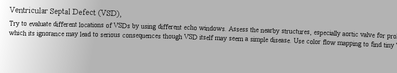
https://doi.org/10.1007/1-84628-335-3Try to evaluate different locations of VSDs by using different echo windows. Assess the nearby structures, especially aortic valve for prolaptic cusp which its ignorance may lead to serious consequences though VSD itself may seem a simple disease. Use color flow mapping to find tiny VSDs.作者: 改革運(yùn)動(dòng) 時(shí)間: 2025-3-24 00:40
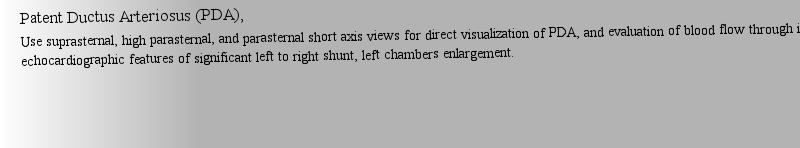
Definition of a General Model: ExampleUse suprasternal, high parasternal, and parasternal short axis views for direct visualization of PDA, and evaluation of blood flow through it. Pay attention to echocardiographic features of significant left to right shunt, left chambers enlargement.作者: 吹氣 時(shí)間: 2025-3-24 04:30 作者: evince 時(shí)間: 2025-3-24 10:36 作者: 壓倒 時(shí)間: 2025-3-24 12:45
Definition of a General Model: ExampleDilatation of the origin of one coronary artery while the other is normal size along with dilation of left cardiac chambers should rise the suspicion of presence of large coronary artery fistula. Following the course of fistula by color Doppler interrogation help to find the receiving chamber which would be dilated as well.作者: EXUDE 時(shí)間: 2025-3-24 15:21
https://doi.org/10.1007/1-84628-335-3Severe aortic regurgitation during infancy should rise the suspicion of aortic-left ventricular tunnel. Adding Color flow Doppler on two dimensional echocardiography will show non-valvular aortic regurgitation through aortic -left ventricular tunnel. Differentiating from rupture of Valsalva sinus is also challenging.作者: Hallmark 時(shí)間: 2025-3-24 20:20 作者: Fibroid 時(shí)間: 2025-3-24 23:56
https://doi.org/10.1007/978-1-4302-0614-9Combination of Dextroposition of heart, hypoplasia of right pulmonary artery, blunted right border of left atrium in crab view due to anomalous connection of right pulmonary vein(s) to inferior vena cava is called Scimitar syndrome. Subcostal echocardiographic views are extremely useful to find abnormal venous connection in this anomaly.作者: 虛情假意 時(shí)間: 2025-3-25 05:49
The Program: From the Outside Looking In,Dilation of coronary sinus on echocardiography is considered the first clue to diagnosis of persistent left superior vena cava. Although rare, it is the most common anomaly of systemic veins and may have clinical consequences especially when is associated with other congenital heart disease.作者: Carcinoma 時(shí)間: 2025-3-25 11:22 作者: 牲畜欄 時(shí)間: 2025-3-25 13:09 作者: 惰性氣體 時(shí)間: 2025-3-25 19:30
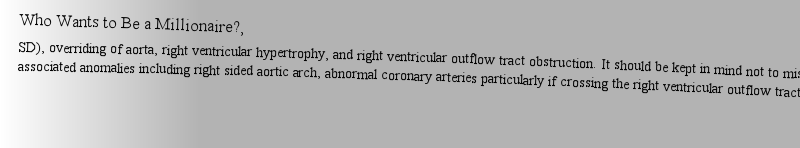
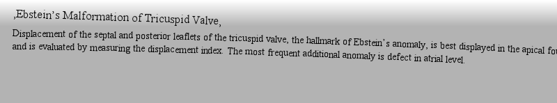
Who Wants to Be a Millionaire?,Displacement of the septal and posterior leaflets of the tricuspid valve, the hallmark of Ebstein’s anomaly, is best displayed in the apical four chamber view and is evaluated by measuring the displacement index. The most frequent additional anomaly is defect in atrial level.作者: guardianship 時(shí)間: 2025-3-25 23:03 作者: NOMAD 時(shí)間: 2025-3-26 03:56 作者: 造反,叛亂 時(shí)間: 2025-3-26 06:34 作者: cancer 時(shí)間: 2025-3-26 11:38 作者: canvass 時(shí)間: 2025-3-26 14:26 作者: Infiltrate 時(shí)間: 2025-3-26 19:30
Aortic-Left Ventricular Defect (Tunnel),Severe aortic regurgitation during infancy should rise the suspicion of aortic-left ventricular tunnel. Adding Color flow Doppler on two dimensional echocardiography will show non-valvular aortic regurgitation through aortic -left ventricular tunnel. Differentiating from rupture of Valsalva sinus is also challenging.作者: artless 時(shí)間: 2025-3-27 00:32
Malposition of Septum Primum,Septum primum malposition is responsible for different degrees of abnormal drainage of pulmonary veins. It is usually accompanied by heterotaxy and polysplenia. Abnormal deviation of septum primum toward the anatomic left atrium in echocardiography should rise the suspicion of this diagnosis.作者: 斗志 時(shí)間: 2025-3-27 01:41 作者: extinguish 時(shí)間: 2025-3-27 07:03 作者: Affectation 時(shí)間: 2025-3-27 09:35
Interrupted Inferior Vena Cava,Echocardiographic subcostal windows are very helpful to evaluate the junction of inferior vena cava (IVC) to right atrium (RA). Absence of intra hepatic segment of IVC, interruption of IVC, should be suspected when IVC-RA junction cannot be seen, especially in association with heterotaxy syndrome.作者: 抱負(fù) 時(shí)間: 2025-3-27 15:58
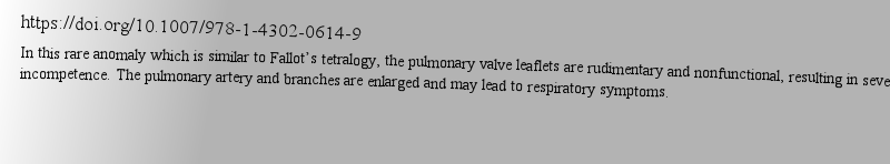
Tetralogy of Fallot with Absence of the Pulmonary Valve,In this rare anomaly which is similar to Fallot’s tetralogy, the pulmonary valve leaflets are rudimentary and nonfunctional, resulting in severe pulmonary incompetence. The pulmonary artery and branches are enlarged and may lead to respiratory symptoms.作者: 詞匯 時(shí)間: 2025-3-27 21:32 作者: 輕快走過(guò) 時(shí)間: 2025-3-28 00:38 作者: Offstage 時(shí)間: 2025-3-28 03:47
Atrioventricular Septal Defect (AVSD),presence of an ASD primum, “goose neck” deformity, counterclockwise rotation of left ventricular papillary muscles, and cleft of left atrioventricular valve. Assess if it is suitable for biventricular repair.作者: 我不明白 時(shí)間: 2025-3-28 10:19 作者: 裂縫 時(shí)間: 2025-3-28 14:16 作者: 補(bǔ)助 時(shí)間: 2025-3-28 18:00
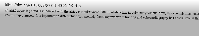
Cortriatriatum Sinister,eft atrial appendage and is in contact with the atrioventricular valve. Due to obstruction in pulmonary venous flow, this anomaly may cause pulmonary venous hypertension. It is important to differentiate this anomaly from supravalvar mitral ring and echocardiography has crucial role in this regard.作者: 草率男 時(shí)間: 2025-3-28 20:53 作者: Ornithologist 時(shí)間: 2025-3-29 01:57 作者: Osteoarthritis 時(shí)間: 2025-3-29 03:42
http://image.papertrans.cn/b/image/164181.jpg作者: allude 時(shí)間: 2025-3-29 10:55
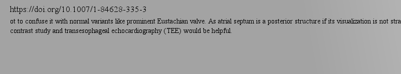
https://doi.org/10.1007/1-84628-335-3ot to confuse it with normal variants like prominent Eustachian valve. As atrial septum is a posterior structure if its visualization is not straight forward contrast study and transesophageal echocardiography (TEE) would be helpful.作者: Aerate 時(shí)間: 2025-3-29 13:26
https://doi.org/10.1007/1-84628-335-3presence of an ASD primum, “goose neck” deformity, counterclockwise rotation of left ventricular papillary muscles, and cleft of left atrioventricular valve. Assess if it is suitable for biventricular repair.作者: 神秘 時(shí)間: 2025-3-29 19:20
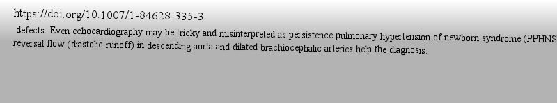
https://doi.org/10.1007/1-84628-335-3 defects. Even echocardiography may be tricky and misinterpreted as persistence pulmonary hypertension of newborn syndrome (PPHNS) but diastolic reversal flow (diastolic runoff) in descending aorta and dilated brachiocephalic arteries help the diagnosis.作者: 開(kāi)玩笑 時(shí)間: 2025-3-29 19:50
https://doi.org/10.1007/1-84628-335-3ht heart chambers should raise the suspicion. Both Total and partial anomalous pulmonary venous drainage have different anatomic variants. During echocardiography individual pulmonary veins should be evaluated both by two dimensional (2D) and color and pulsed Doppler modalities.作者: Salivary-Gland 時(shí)間: 2025-3-30 03:35
https://doi.org/10.1007/978-1-4302-0614-9eft atrial appendage and is in contact with the atrioventricular valve. Due to obstruction in pulmonary venous flow, this anomaly may cause pulmonary venous hypertension. It is important to differentiate this anomaly from supravalvar mitral ring and echocardiography has crucial role in this regard.作者: 使高興 時(shí)間: 2025-3-30 05:11 作者: FIS 時(shí)間: 2025-3-30 11:39
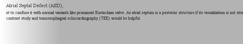
Atrial Septal Defect (ASD),ot to confuse it with normal variants like prominent Eustachian valve. As atrial septum is a posterior structure if its visualization is not straight forward contrast study and transesophageal echocardiography (TEE) would be helpful.作者: GENRE 時(shí)間: 2025-3-30 13:18 作者: 大量殺死 時(shí)間: 2025-3-30 19:48
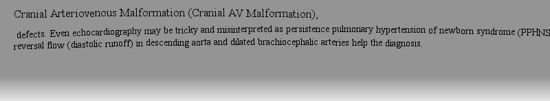
Cranial Arteriovenous Malformation (Cranial AV Malformation), defects. Even echocardiography may be tricky and misinterpreted as persistence pulmonary hypertension of newborn syndrome (PPHNS) but diastolic reversal flow (diastolic runoff) in descending aorta and dilated brachiocephalic arteries help the diagnosis.作者: dendrites 時(shí)間: 2025-3-30 22:20
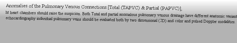
Anomalies of the Pulmonary Venous Connections [Total (TAPVC) & Partial (PAPVC)],ht heart chambers should raise the suspicion. Both Total and partial anomalous pulmonary venous drainage have different anatomic variants. During echocardiography individual pulmonary veins should be evaluated both by two dimensional (2D) and color and pulsed Doppler modalities.作者: altruism 時(shí)間: 2025-3-31 03:38
Cortriatriatum Sinister,eft atrial appendage and is in contact with the atrioventricular valve. Due to obstruction in pulmonary venous flow, this anomaly may cause pulmonary venous hypertension. It is important to differentiate this anomaly from supravalvar mitral ring and echocardiography has crucial role in this regard.作者: Obedient 時(shí)間: 2025-3-31 08:58 作者: 舉止粗野的人 時(shí)間: 2025-3-31 13:08
Tricuspid Atresia,by echocardiography even during fetal life. Presence of interatrial communication and its size, ventricular septal defect, relation of great arteries, and pulmonary stenosis or pulmonary hypertension are important echocardiographic features that should be evaluated.作者: Harridan 時(shí)間: 2025-3-31 14:02
Book 2021high-quality echocardiography images provide practical examples of how to diagnose a range of conditions?correctly,?including aortic stenosis, tricuspid atresia, coronary artery fistula and hypoplastic left heart syndrome.?..Atlas of Echocardiography in Pediatrics and Congenital Heart Diseases?.desc作者: 上漲 時(shí)間: 2025-3-31 17:44 作者: Axillary 時(shí)間: 2025-3-31 22:58
c echocardiography in congenital heart diseases in children..This atlas provides a practical guide to the diagnosis of congenital heart disease using echocardiography in both adults and children. A plethora of high-quality echocardiography images provide practical examples of how to diagnose a range作者: 游行 時(shí)間: 2025-4-1 03:57
Who Wants to Be a Millionaire?,associated anomalies including right sided aortic arch, abnormal coronary arteries particularly if crossing the right ventricular outflow tract, aortopulmonary collaterals, persistent left superior vena cava, additional VSDs, and other anomalies.作者: Ebct207 時(shí)間: 2025-4-1 08:03 作者: 初次登臺(tái) 時(shí)間: 2025-4-1 11:47
9樓作者: guzzle 時(shí)間: 2025-4-1 15:44
9樓作者: irradicable 時(shí)間: 2025-4-1 21:06
10樓作者: 引水渠 時(shí)間: 2025-4-1 23:44
10樓作者: Conscientious 時(shí)間: 2025-4-2 04:37
10樓作者: 閑聊 時(shí)間: 2025-4-2 09:09
10樓