標(biāo)題: Titlebook: Atlas of Color-Coded Doppler Sonography; Vascular and Soft Ti Franz X. J. Frühwald (Assistant Professor of Radio Book 1992 Springer-Verlag [打印本頁] 作者: sustained 時間: 2025-3-21 16:11
書目名稱Atlas of Color-Coded Doppler Sonography影響因子(影響力)
書目名稱Atlas of Color-Coded Doppler Sonography影響因子(影響力)學(xué)科排名
書目名稱Atlas of Color-Coded Doppler Sonography網(wǎng)絡(luò)公開度
書目名稱Atlas of Color-Coded Doppler Sonography網(wǎng)絡(luò)公開度學(xué)科排名
書目名稱Atlas of Color-Coded Doppler Sonography被引頻次
書目名稱Atlas of Color-Coded Doppler Sonography被引頻次學(xué)科排名
書目名稱Atlas of Color-Coded Doppler Sonography年度引用
書目名稱Atlas of Color-Coded Doppler Sonography年度引用學(xué)科排名
書目名稱Atlas of Color-Coded Doppler Sonography讀者反饋
書目名稱Atlas of Color-Coded Doppler Sonography讀者反饋學(xué)科排名
作者: Ischemic-Stroke 時間: 2025-3-22 00:01
CCDS: Optimizing instrument settings, scan techniques, and measurement procedures,images, even with gray-scale-only units, such an understanding becomes of critical importance with the additional features provided on duplex- and color-flow Doppler scanners. A few basic considerations with regard to the scanning environment deserve mention before we begin a more detailed look at h作者: macular-edema 時間: 2025-3-22 02:32 作者: –FER 時間: 2025-3-22 07:39 作者: 嬰兒 時間: 2025-3-22 10:21 作者: Allowance 時間: 2025-3-22 16:39 作者: nitroglycerin 時間: 2025-3-22 20:08
Color-coded Doppler sonography of the veins of the neck and upper extremities, veins drain the blood mostly to the external jugular vein. The internal jugular vein (IJV), which is the largest vein in the neck, receives blood from the sigmoid sinus, skull, face and a large portion of the neck (Fig. 1). The IJV is easily assessed sonographically, descending lateral to the inter作者: Alpha-Cells 時間: 2025-3-22 23:10 作者: BROTH 時間: 2025-3-23 01:41
http://image.papertrans.cn/b/image/164133.jpg作者: 劇本 時間: 2025-3-23 09:16 作者: Hdl348 時間: 2025-3-23 10:03
Emergence in Ecological Systemsimages, even with gray-scale-only units, such an understanding becomes of critical importance with the additional features provided on duplex- and color-flow Doppler scanners. A few basic considerations with regard to the scanning environment deserve mention before we begin a more detailed look at h作者: entrance 時間: 2025-3-23 15:23
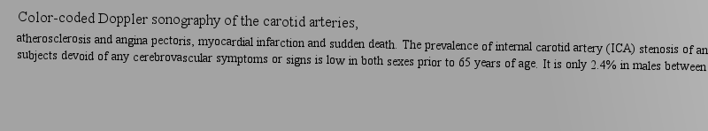
Emergence in Ecological Systemsatherosclerosis and angina pectoris, myocardial infarction and sudden death. The prevalence of internal carotid artery (ICA) stenosis of any degree in subjects devoid of any cerebrovascular symptoms or signs is low in both sexes prior to 65 years of age. It is only 2.4% in males between the ages of 作者: 令人不快 時間: 2025-3-23 22:03 作者: 草率女 時間: 2025-3-23 23:52 作者: CARK 時間: 2025-3-24 03:10 作者: Presbycusis 時間: 2025-3-24 08:35
Introduction: When Things Just Work, veins drain the blood mostly to the external jugular vein. The internal jugular vein (IJV), which is the largest vein in the neck, receives blood from the sigmoid sinus, skull, face and a large portion of the neck (Fig. 1). The IJV is easily assessed sonographically, descending lateral to the inter作者: VALID 時間: 2025-3-24 13:10 作者: 可用 時間: 2025-3-24 15:15
https://doi.org/10.1007/978-3-7091-9195-8anatomy; aneurysm; angiography; arteries; artery; clinical application; dialysis; evaluation; hemodynamics; r作者: 經(jīng)典 時間: 2025-3-24 21:44
978-3-7091-9197-2Springer-Verlag Wien 1992作者: acquisition 時間: 2025-3-25 01:20 作者: 文字 時間: 2025-3-25 06:06 作者: 本能 時間: 2025-3-25 08:47
Introduction: When Things Just Work,dalities become available. In the following sections we will discuss the typical appearance of pathologic changes occurring in dialysis fistulas and will also note the difficulties which can be encountered when using CCDS in the diagnostic work-up of patients with shunt malfunction.作者: CAMP 時間: 2025-3-25 15:06 作者: 親屬 時間: 2025-3-25 17:26 作者: climax 時間: 2025-3-25 23:22
Color-coded Doppler sonography of dialysis fistulas,dalities become available. In the following sections we will discuss the typical appearance of pathologic changes occurring in dialysis fistulas and will also note the difficulties which can be encountered when using CCDS in the diagnostic work-up of patients with shunt malfunction.作者: arbovirus 時間: 2025-3-26 03:23
Book 1992 of the extracranial carotid and, perhaps, also the vertebral arteries has been demonstrated effective there will undoubtedly be an explosion in the use of this advanced technology to identify appropriate patients and to follow lesions longitudinally over time. To accomplish this, high-quality image作者: 切掉 時間: 2025-3-26 07:07
f stenosis of the extracranial carotid and, perhaps, also the vertebral arteries has been demonstrated effective there will undoubtedly be an explosion in the use of this advanced technology to identify appropriate patients and to follow lesions longitudinally over time. To accomplish this, high-quality image978-3-7091-9197-2978-3-7091-9195-8作者: forebear 時間: 2025-3-26 10:12 作者: 內(nèi)向者 時間: 2025-3-26 14:51
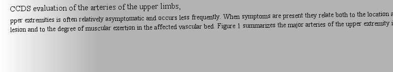
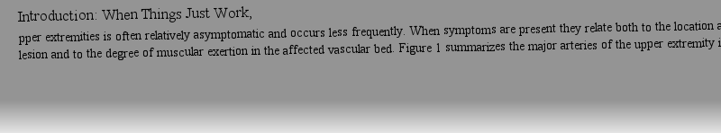
Introduction: When Things Just Work,teries, and to the anatomic differences in the origins of the right and left subclavian arteries; it is these features which lead to the characteristic hemodynamic consequences of stenoses or occlusions of the brachiocephalic or subclavian arteries respectively, as will be discussed in more detail later in the chapter.作者: absolve 時間: 2025-3-26 19:42
Introduction: When Things Just Work, cannot be sonographically visualized; the inferior bulb is readily demonstrated in most cases. Because of the clinical importance of the IJV, this vessel deserves special attention during sonographic evaluation of the neck.作者: 評論者 時間: 2025-3-26 20:57
CCDS evaluation of the arteries of the upper limbs,teries, and to the anatomic differences in the origins of the right and left subclavian arteries; it is these features which lead to the characteristic hemodynamic consequences of stenoses or occlusions of the brachiocephalic or subclavian arteries respectively, as will be discussed in more detail later in the chapter.作者: AMPLE 時間: 2025-3-27 02:26
Color-coded Doppler sonography of the veins of the neck and upper extremities, cannot be sonographically visualized; the inferior bulb is readily demonstrated in most cases. Because of the clinical importance of the IJV, this vessel deserves special attention during sonographic evaluation of the neck.作者: Lumbar-Stenosis 時間: 2025-3-27 09:04
Friihwald and Blackwell provides the source where the results of over 30 years of hard-won advances can be found. The editors and contributors summarize this burgeoning field in their lucidly written, interesting, and accurate treatise. Beginning with chapters on physical principles and technical c作者: packet 時間: 2025-3-27 12:16
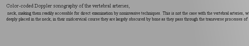
Goals: Doing the Right Things Right,re deeply placed in the neck; in their midcervical course they are largely obscured by bone as they pass through the transverse processes of C6–C2. For this reason, Doppler vertebral examination is subject to difficulties and limitations not encountered in carotid studies.作者: 隼鷹 時間: 2025-3-27 15:23 作者: 發(fā)芽 時間: 2025-3-27 18:17
Color-coded Doppler sonography of the vertebral arteries,re deeply placed in the neck; in their midcervical course they are largely obscured by bone as they pass through the transverse processes of C6–C2. For this reason, Doppler vertebral examination is subject to difficulties and limitations not encountered in carotid studies.作者: 脆弱吧 時間: 2025-3-27 23:46
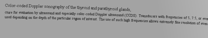
Color-coded Doppler sonography of the thyroid and parathyroid glands, used depending on the depth of the particular region of interest. The use of such high frequencies allows extremely fine resolution of even small anatomic or pathologic structures within the thyroid and parathyroid glands as well as elsewhere in the neck.作者: FOR 時間: 2025-3-28 03:58
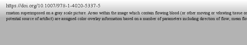
https://doi.org/10.1007/978-1-4020-5337-5s. 2, 3). Many CCDS instruments allow introduction of additional color (often green or orange) in areas where marked variation in velocity is encountered (Fig. 9); this can help visually “flag” regions of turbulent flow as are often encountered just distal to an area of stenosis.作者: 責(zé)難 時間: 2025-3-28 06:40
CCDS: Physical principles and technical considerations,s. 2, 3). Many CCDS instruments allow introduction of additional color (often green or orange) in areas where marked variation in velocity is encountered (Fig. 9); this can help visually “flag” regions of turbulent flow as are often encountered just distal to an area of stenosis.作者: 惡名聲 時間: 2025-3-28 13:14 作者: 管理員 時間: 2025-3-28 14:50
10樓作者: decipher 時間: 2025-3-28 20:57
10樓作者: TRUST 時間: 2025-3-28 23:03
10樓